  |
  |
Primary Vascular Tissue

These images are superb examples of the eustele arrangement of primary vascular bundles. The above image is a t-blue stained cross-section which clearly shows the pith, cortex, and interfascicular parenchyma surrounding the vascular bundles. The phloem is closer to the surface of the stem than the xylem (the vessel members are clearly evident in the xylem).

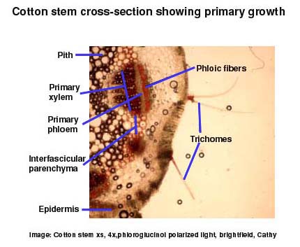
Overall primary growth can be nicely summarized in the following image which shows the elements discussed above in a phloroglucinol stained cross-section. 
To read about primary growth in the ground tissue system... |

Introduction Flowers&Fruit Roots Stems Leaves |