To avoid major economic losses, those diseases of greater frequency and severity require the most attention to diagnosis and control. Sometimes symptom expression may vary because of factors such as environment and variety. New diseases may develop as conditions and varieties change.
When such variations occur, collect samples and submit them through the county Extension agent for further microscopic examination and analysis.
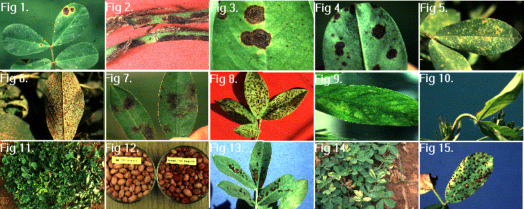
                                   EARLY LEAF SPOT          LATE LEAF SPOT

 Seasonal development                 Any Time           Usually late season

 Shape of spot                  Circular to irregular      Usually circular

 Yellow halo                       Usually (Fig.1)             Sometimes

 Stem lesions                       Yes (Fig. 2)                  Yes

 Leaf surface where                    Upper                     Lower
 most spore form

 Appearance of upper              Brown and slightly        Brown to black
 leaf surface                     raised (Fig. 3)           and smooth

 Appearance of lower              Brown and usually         Black and rough
 leaf surface                     smooth                    or granular (Fig. 4)
Rust first appears as a yellowish-green fleck on the upper leaf surface. Almost simultaneously pustules appear as small raised bumps on the lower leaf surface. Certain other peanut diseases may be improperly identified as rust. Figure 5.
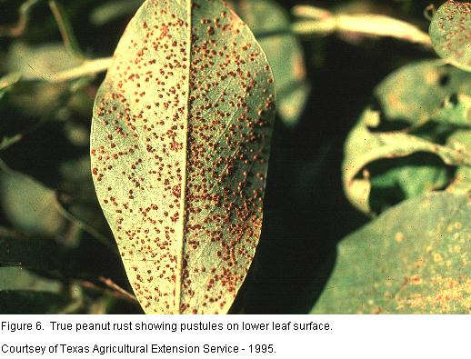
True peanut rust always has pustules filled with spores on the leaf surface. Rubbing a white tissue or handkerchief across these pustules reveals the presence of reddish-brown spores. Figure 6.
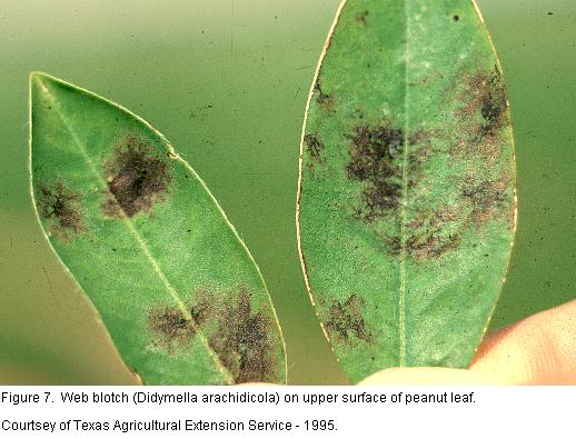
Webbing or netting patterns produced by the fungus on the upper surface of peanut leaves are the most distinct symptoms. Webbing occurs when fungal strands grow just beneath the waxy cuticle on the leaf surface. These strands tend to grow together and form a blotch that almost resembles a fingerprint on the leaf. Sometimes only webbing occurs, and no blotch is formed. This disease is most prevalent of Spanish type peanuts. Figure 7.
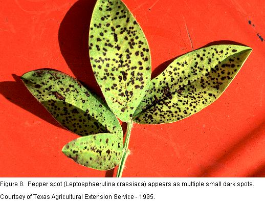
Pepper spot usually appears as multiple small dark brown to black specks on the leaf surface. These specks gradually enlarge but do not reach the size of the more common early leaf spot. Spots grow together to form larger spots under the most severe disease pressure. Pepper spot has never been acknowledged as a serious peanut disease. The fungus is considered a weak pathogen. Figure 8
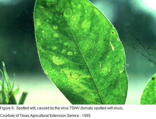
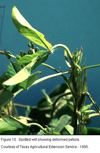
This disease occurs in many parts of Texas but major yield loss has been limited to the peanut growing areas of South Texas. Certain thrip species transmit the tomato spotted wilt virus. Young plants show ringspots (Figure 9), a yellow-green mottle pattern, brown spots and ringspots, or combinations of these. The first or second leaf on each terminal that develops sysmptoms often has a twisted and downward curved petiole (Figure 10).
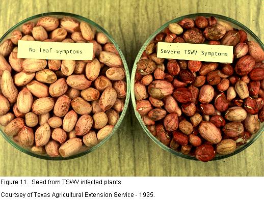
Lower leaves of young infected plants remain green. Plants infected early in the season become stunted with bunched growth and small deformed leaves (Figure 11). Late in the season, newly infected plants are more likely to turn yellow, wilt and die as leaf growth slows and stops. Plants with systemic yellowing symptoms often have internal taproot discoloration. Plants infected early in the season produce distorted, fewer and smaller pods than healthy plants. Seed from infected plants may be dark red (Figure 12) or have brown and white streaks. The virus is not considered to be seedborne, however.
Ozone may be produced during electrical storms or may be brought down from the upper atmosphere by strong down drafts. A scorced appearance occurs primarily on the upper leaf surface. Damage is usually evident in 4-5 days, and scorced leaves soon drop. Spanish varities are much more susceptible thatn Runner varieties. Figure 13.
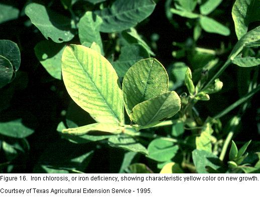
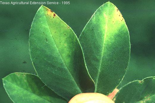
Peanuts grown in alkaline soils often display a characteristic yellow color on the new growth (Figure 16). Veins remain green, and the yellow color develops between veins. Severe deficiency may cause leaves to be almost white. Deficiencies of other micronutrients such as zinc and copper may produce a similar condition in peanuts. Chemical analysis of the plant tissue is the only sure way to differentiate among minor element deficiencies.
This insect feeds along the mid rib producing yellow leaf tips. The tip of the leaflet yellows and tips or "beaks" downward. Heavy feeding damage gives a yellow appearance to the field. Figure 17
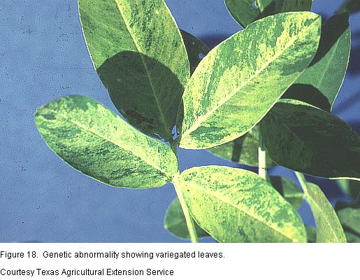
Variegated leaves are often found in fields of all types of peanuts. This symptom may be present only on leaves from one branch of a plant with all other branches displaying normal leaves. This condition is thought to be genetic in origin, and no organism is involved. The green chlorophyll pigment fails to develop and the yellowed variegation occurs. Figure 18
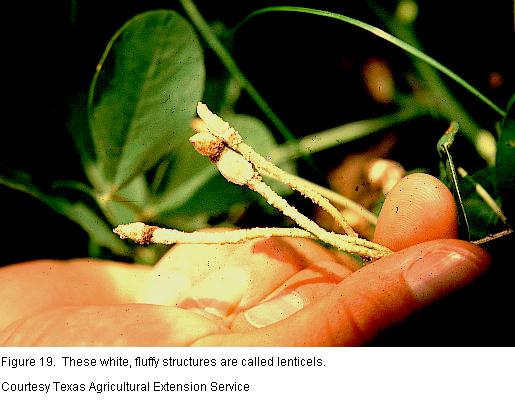
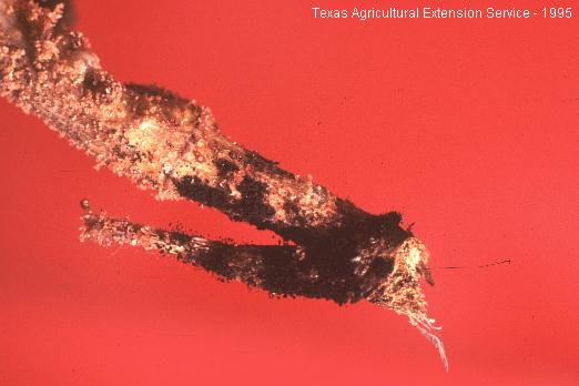
Enlarged white fluffy structures called lenticels form on pegs and pods when soil is saturated with water. This is the means by which surface area is increased to contact more available oxygen. Lenticels are localized enlargments of plant cells. Figure 19
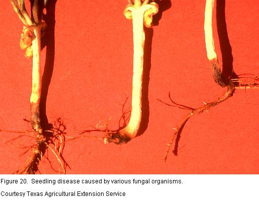
Soil borne organisms attack peanut seedlings at various stages in their development. Poor quality, low energy or physically damaged seed may actually rot before germination. Plants from healthy seed that germinate well may develop brown to black spots on the elongating below-ground parts. Under cold or unfavorable soil conditions these lesions increase rapidly and often girdle and kill the seedling before it can become established. The problem is more severe in some fields than others. Later planting in warmer soils tends to reduce the potential for seedling disease. Figure 20
These fungi produce a chemical toxin called aflatoxin, and infected peanuts are placed in Segregation III at the buying point. These fungi produce masses of yellow green spores. Slight magnification of pods and kernels show fluffy ball-like structures on tiny stalks. The occurrence of Segregation III peanuts is usually associated with drought stress or high moistaure harves conditions.Figure 21
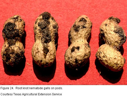
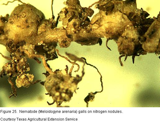
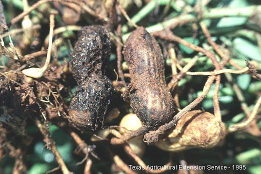
Root knot nematodes cause gall formation on roots (Figure 22), pegs (Figure 23), pods (Figure 24) and nitrogen nodules (Figure 25). Nematode-induced galls are formed in the root and pod tissue and are not attached to the side of the root as is the case with nitrogen nodules. Severe yellowing and stunting of the plant and even premature death are all symptoms of this disease. Nematode numbers are highest when soil, pod and root tissue are sampled just prior to harvest.
Root lesion nematodes invade and feed on below-ground peanut plant parts and can best be identified by the presence on pods of small tan spots with dark centers. Feedling damage permits other organisms to enter. Weakened pegs commonly cause pods to drop from the plant. Figure 26.
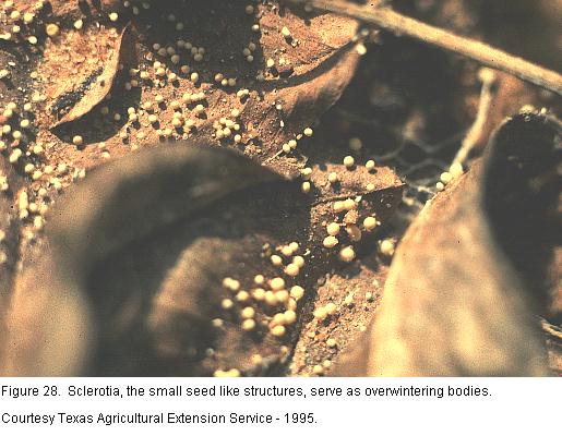
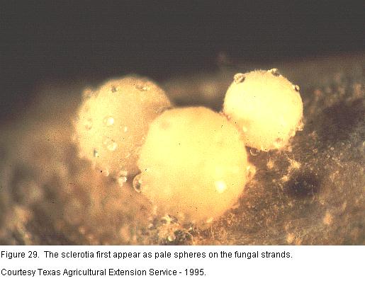
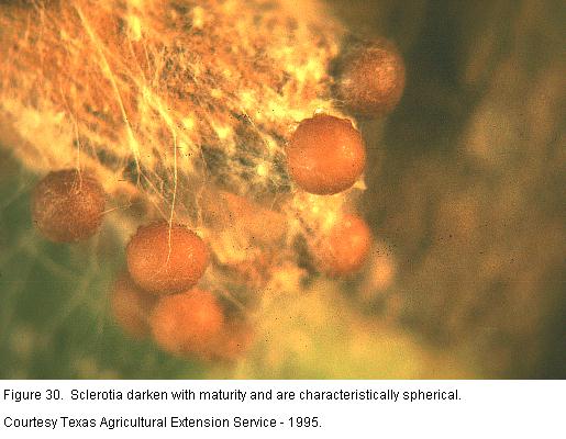
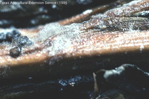
During periods of warm, moist weather this fungus develops as a mass of white fungal strands (Figure 27) at or near the soil surface. The fungus can grow directly on peanut vines and also forms on fallen leaves. As the fungus matures it produces small, seedlike structures call sclerotia (Figure 28) that serve as overwintering bodies. The sclerotia first appear as tiny white dots on the fungal strands and darken with maturity (Figure 29). The distinguishing characteristic of this fungus is the almost perfect spherical shape of it sclerotia (Figure 30).
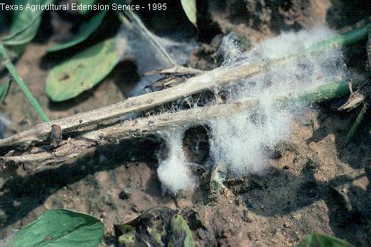
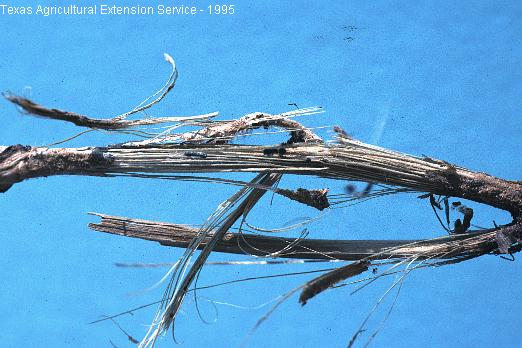
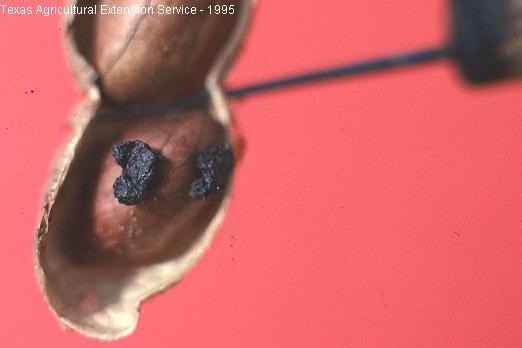
Early symptoms of this fungal disease include small white tufts of cotton-like growth on the stems at leaf axils near the ground line (Figure 31). Recent stem damage is light tan in color. Later stages of the disease show up as severe stem shredding (Figure 32) almost as if the stem had exploded, accompanied by the production of many small black irregularly shaped slerotia. The fungus grows most rapidly at 26 degree C (79 degrees F). Sclerotia are produced most rapidly at 22 degrees C (72 degrees F) on all plant parts, in soil, and inside limb and pod tissue (Figure 33).
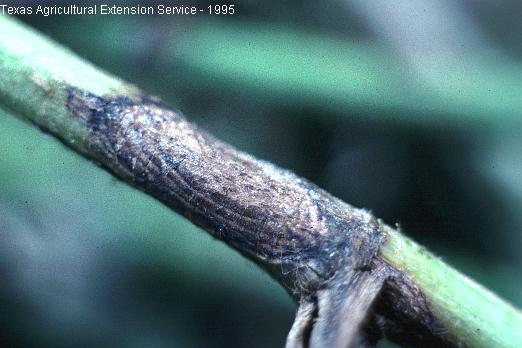
This common disease occurs in all peanut growing areas of Texas. Lesions or spots produced by this fungus on limbs (Figure 34), pegs and pods (Figure 35) are brown to black and slightly sunken. A series of concentric rings often appears on limb lesions. Rhizoctonia rot is much more likely to be a problem where peanuts have been grown continuously. Fields in areas never before planted to peanuts may, however, experience significant losses to this organism.
Pods affected by this fungus are initially light brown in color and turn dark brown with age. Infected tissue is watery when squeezed. The disease is more prevalent under wet field conditions which favor development of this "water mold" fungus. Field symptoms may not always be reliable for accurate diagnosis since other organisms are almost always present in the diseased tissue. Figure 36.
This fungal disease occurs predominantly in certain areas of West Texas. Symptoms of this disease closely resemble Sclerotinia blight. The fluffy mycelial growth has a slight gray tint, and the sclerotia are almost twice the size of those produce by the Sclerotinia blight fungus. Microscopic observation of fungal structures is often required to distinguish between the two diseases. Figure 37.
Late planting, insect feeding, high soil temperature and drought stress for the first few weeks after planting have all been associated with the incidence of this problem. Death of plants can occur at any stage from seedling to harvest. The fungus attacks the crown or collar near the soil line and soon girdles and kills the plant. The slightly fluffy, black appearance of fungal fruiting structures at the ground line is the best field diagnostic symptom. Figure 38.
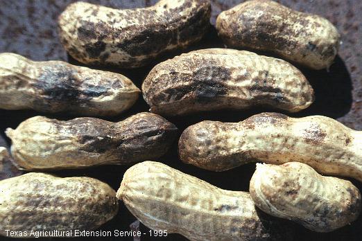
Loss of natural pod color is the primary problem caused by this fungal organism. The external layer of pod tissue turns dark brown to black but the kernal is seldom damaged. There is consequently much greater concern about peanuts grown for the "in shell" trade. This disease is found primarily in the Texas peanut producing counties bordering New Mexico. Figure 39.
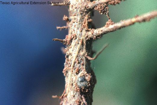
Affected plants die suddenly, and leaves remain firmly attached. Plants may be easily pulled from the soil because root tissue decays rapidly. Buff to brown strands of the fungus can be observed on the surface of larger roots. The disease is more prevalent in alkaline soils. Figure 40.
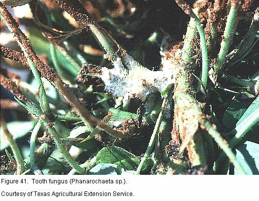
This fungus often occurs in peanut fields that have been rotated with cotton. The fleshy white fungus, with what appears to be tiny, sharp, protruding teeth, is not a known pathogen of peanuts. It is associated with old woody cotton stalk debris. Some may confuse its development with certain other pathogens of peanuts. Figure 41.
Plant disease symptoms not fully described in this publication may
be occasionally encountered. Contact your county Extension agent for
additional diagnostic assistance.
New or unusual diseases that occur may require laboratory diagnosis. Samples needing such attention may be sent to:
Texas Plant Disease Diagnostic Laboratory
Room 101, L.F. Peterson Building
Texas A&M University
College Station, TX 77843-2132
409-845-8032